| Quantity | 3+ units | 10+ units | 30+ units | 50+ units | More |
|---|---|---|---|---|---|
| Price /Unit | $2,004.48 | $1,963.57 | $1,902.21 | $1,820.40 | Contact US |
 RS-N60 Full Digital PC Processing System Veterinary Ultrasonic Diagnostic Instrument 128G SSD 15-inch LCD
$1,885.99
RS-N60 Full Digital PC Processing System Veterinary Ultrasonic Diagnostic Instrument 128G SSD 15-inch LCD
$1,885.99
 T6 Cattle Full Digital Ultrasonic Diagnostic Instrument 7-inch Touch Screen with Rectal Linear Array Probe
$1,915.14
T6 Cattle Full Digital Ultrasonic Diagnostic Instrument 7-inch Touch Screen with Rectal Linear Array Probe
$1,915.14
 M6 Mechanical Fan Scan Veterinary Ultrasonic Diagnostic Instrument Support Probe Preheating with 5.7-inch UHD LCD
$528.10
M6 Mechanical Fan Scan Veterinary Ultrasonic Diagnostic Instrument Support Probe Preheating with 5.7-inch UHD LCD
$528.10
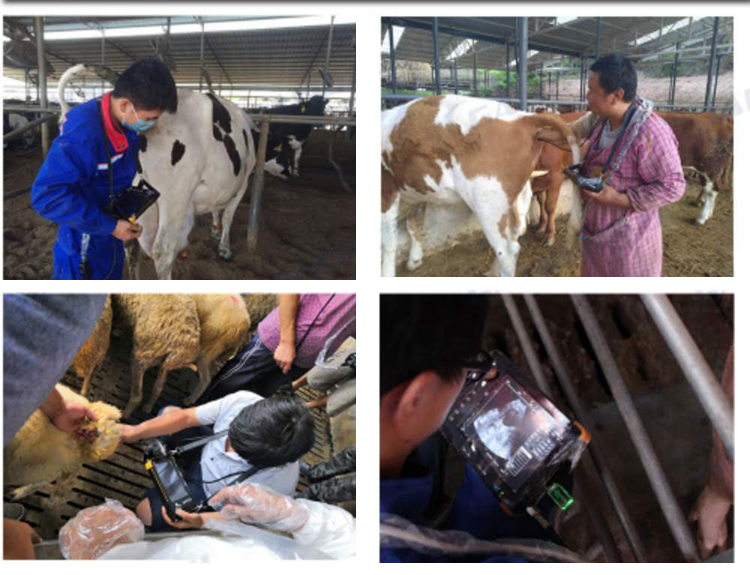
Portable Veterinary Ultrasound Scanner 7 Inch LCD Screen for Large Animals Cow Horse Donkey GDF-K8
Description:
Our DGF-K8 is equipped with 6.5MHz linear rectal probe, which is suitable for large animals including horses, cows and donkeys. It is widely used in in pasture, animal hospitals, veterinary stations and clinical teaching. It can be used to detect early pregnancy, follicle monitoring, fetal vitality, uterine disease, etc.
Main Functions:
- For pregnant animals
- Master postpartum uterus recovery
- Diagnosis of uterine diseases
- Observing embryo development
- Measuring fetal heart rate
- Estimate the number of babies
Features:
- Language: English
- Waterproof device
- 7 Inch full digital LCD display screen
- 8G memory to store countless videos and pictures
- 6.5MHz Linear Rectal Probe: perfect for cows, horse and other large animals
- Longer Working Time: It can be recharged in just 6hours and works for 3 hours
- Super long standby time
Specifications:
- Model: GDF-K8
- Probe: 6.5MHz Frequency-Conversion Electronic Rectal Probe
- Effective Depth: ≥50mm
- Dead Zone: ≤3mm
- Resolution:
Horizontal direction: ≤1mm (Depth: ≤ 60mm)
Vertical direction: ≤0.5mm (Depth: ≤ 50mm)
- Geometric Position Accuracy: Horizontal≤5%; Vertical≤5%
- Image Magnification: x1.0, x1.2, x1.5, x2.0
- Scanning Way: Electronic Linear Array
- Working Frequency: 2.0MHz-10MHz
- Display Modes: B, B+B, B+M, M, 4B
- Monitor Size: 7.0 Inch LCD Screen
- Focus Adjustment: number of focus, focus position
- Body Landmark: 8 Types
- Image Processing: pseudocolor, grayscale correction, image smoothing, histogram
- Frequency Adjustment: 3 Levels
- Frame Related Adjustment: Support
- Scanning Angle: visually adjustable
- Measurement: distance, perimeter, area, volume, gestational age, expected delivery date
- Characters & Notes: date, clock, name, PID, age, gender, etc.
- Image Grayscale: 256 levels
- Number of Scan Lines: 512 lines/frame
- Frame Rate: 30 frames / sec
- Digital Scan Converter: 512 × 512 × 8bits
- Machine size: 228mm × 152mm × 37mm
- Machine weight: 981g
- Memory Capacity: up to 8GB
Package Included:
- 1 x Main Unit
- 1 x 6.5MHz Linear Rectal Probe
- 1 x User Manual
- 1 x Power Adapter
- 1 x Carry Case
Detection Method for Cows:
1. Selection of Position: The uterus of the cow in early pregnancy is located before and after the entrance of the pelvic cavity. The best part of the exploration from the outside of the body is occupied by a large breast in front of the pubic bone. The left side is the rumen, the right side is the intestinal tract, and the buttocks are thick. The muscle groups are far from the uterus in early pregnancy. Therefore, the best exploration site can only be the vaginal or rectum near the ion palace. In the second and third trimesters, when the uterus sag to the abdominal wall, it can be explored in the lateral lower abdominal wall.
2. Testing Method: Cows take natural standing postures and is fixed on the cow bed or the column. Just like the rectal palpation and the rectum grasping the insemination, one person can operate without special protection. The assistant can help fix the tail of the animal if necessary.
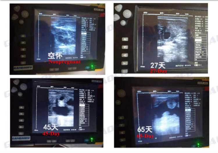
Intravaginal Detection: One hand separates the vulva, and the other hand sends the sterilized long-rod vaginal probe into the vulva, and slowly sends it to the deep part of the vagina and reaches the fornix part. A type instrument probes the uterus with fetal water. The D-type instrument probes the blood flow of the uterine artery anastomosis at the dome below the cervix, which is equivalent to 3-5 points and 7-9 points of the clock. At the cervix, the blood of the palace is not detected, and the upper sides of the cervix are generally not detected. Sometimes the fetal blood sound or fetal heart sound can be found under the sides of the cervix. The probe is usually sterilized using a new clean solution. Coupling agents for intravaginal probing should be sterilized, but are generally not required. The probes for vaginal exploration are small, and the cows have no feeling of discomfort.
Intra-rectal Detection: First use a long cup rectal probe to stimulate the cow's anus, promote the defecation of the cow, then send the probe into the anus, and then slowly send it to the deep rectum. D-type instrument probe, when the probe is fed, the wafer surface is first upward, after the lumbar support joint, the fan surface is turned to the left or right side to probe the middle uterine artery, the depth is generally 30-50 cm. Depths varies due to their body type and pregnancy month. The wafer is probed down to the sides, and after 50 days of gestation, it is possible to detect fetal blood, fetal heart sounds or fetal movements. When type A instrument is used , the probe chip faces down, and reaches the front and rear of the pelvic inlet, and scans downward at a 45 degree angle.


 Portable Wearable Head Mounted Display 0.39-inch OLED for Safety Monitoring FPV")


